


매일미사
우리들의 묵상/체험
| 제목 | 21 02 04 목 평화방송 미사 침의 분비 다스려주지 않고 코의 부비동의 나비 굴을 열어 신경계만 다스리자 귀와 대장 안의 반응이 없어 침 분비 중요 인식 | |||
|---|---|---|---|---|
작성자한영구

|
작성일2021-08-02 | 조회수3,748 | 추천수0 |
반대(1)
 신고
신고
|
|
성부와 성자와 성령의 이름으로 아멘. 흠숭 하올 우리 주 성체 예수 그리스도님, 오늘 2월 4일 연중 제4주간 목요일 평화방송 오전 11시 45분을 선택하여 미사에 참례하였습니다. 미사가 시작되자 저의 두 눈 사이 콧날 양쪽 경사 끝 뼈에 머물러 만져주십니다. 두정엽 정수리 가운데에서 약간 오른쪽 위치에 머물러 만져주시면서 빛의 생명을 아래로 주십니다. 두 눈 사이 콧날 끝 뼈에서 저의 생명을 점검하시면서, 질서에 따라 죄의 상처가 있는 곳에 빛의 생명을 주신다고 상상합니다. 이어서 바로 저의 물렁 코뼈 오른쪽 경사 끝 뼈가 있는 곳에 머물러 만져주시면서 두정엽 정수리에서 빛의 생명을 주시어 다스려주십니다. 이어서 저의 오른쪽 눈썹의 왼쪽 부위에 머물러 물렁 코뼈 오른쪽 경사 끝 뼈와 연결하여 빛의 생명을 주시어 다스려주십니다. 그리고 그 두 곳에 머물러 만져주시면서 다스려주시다가 저의 두정엽 정수리 가운데에 머물러 만져주시면서 빛의 생명을 주십니다. 3곳을 연결하여 빛의 생명을 주시어 다스려주십니다. 귀 안에서 내이의 조직의 기관도 움직이는 소리가 들리지 않고 그 3곳에만 머물러 만져주십니다. 그러자 이번에는 저의 전두엽 우뇌 오른쪽 앞이마 살갗 좌우 상하 가운데 위치에 머물러 넓고 가볍게 만져주시면서, 왼쪽도 대칭의 위치인 전두엽 좌뇌 왼쪽 살갗 이마 그 가운데에 가볍게 넓게 만져주십니다. 두 곳을 대칭으로 만져주십니다. 처음 경험하는 것입니다. 잠깐 만져주시고 저의 코의 인두 또는 인두가 아닌 물렁 코뼈 오른쪽 경사 끝 뼈에 머물러 오른쪽 눈썹 왼쪽 부위에 머물러 만져주시면서 이 두 곳을 연결하여 다스려주십니다. 요사이 코의 인두에 머물러 침의 물질대사를 다스려주시어 거의 매번 침을 귀 안의 조직의 기관 안으로 흘러들게 하여 기관이 활발하게 움직이어 청력을 회복시켜 주시었습니다. 코의 인두 위치는 물렁 코뼈 콧날 오른쪽 경사 끝 뼈와 겹치므로, 이 끝 뼈에서 위턱뼈 굴을 열어 나비 굴과 연결하여 나비 굴에 신경계를 다스려주신 것으로 상상홥니다. 그리고 오른쪽 눈썹 왼쪽 부위는 코의 부비동 이마뼈 굴 안에 있는 위치이므로 이마뼈 굴을 열어 나비 굴과 연결하여 신경계인 교감신경 부교감신경 말초신경을 다스려주신다고 상상합니다.신경계를 집중적으로 다스려주신 것으로 상상합니다. ‘영성체기도’에서 저의 심장박동 안에서 창조주 성부 하느님의 심장박동이 고동쳐주시어 그 고동이 저의 허파 윗부분 두 곳 양쪽 그 박동 안에 고동이 퍼져나가 심장과 허파의 가로 두 곳이 뻐근함을 느낍니다. 새 생명을 주시기 시작하십니다. 그러므로 심장 허파 두 곳 이 3곳과 코의 인두 4곳과 오른쪽 눈썹 왼쪽 끝 부위 5곳과 두정엽 정수리 가운데 6곳에 동시에 머물러 만져주시면서 빛의 생명도 주시고 새 생명도 주시어 다스려주십니다. 그리고 이어서 저의 물렁 코뼈 오른쪽 경사 끝 뼈와 오른쪽 눈썹 왼쪽 부위 머물러 연결하여 만져주시고, 저의 오른쪽 코의 인두에서 위로 수직으로 일자로 오른쪽 앞이마 아래 끝부분과 연결하여 만져주십니다. 그리고 전두엽 우뇌 가운데에 머물러 만져주십니다. 그 3곳에 머물러 만져주시어 빛의 생명을 주시어 다스려주십니다. 심장박동도 고동쳐주시므로 심장과 허파의 윗부분 3곳과 머리 6곳에 머물러 만져주시어 다스려주십니다. 귀 안에서는 아무런 움직임이 없습니다. 그 6곳에만 머물러 만져주십니다. 코의 인두가 있는 곳은 물렁 코뼈 오른쪽 경사 끝 뼈와 겹치는 곳입니다. 그리고 이 끝 뼈에서는 위턱뼈 굴과 연결이 되어 열어주시어 나비 굴과 신경계를 다스려주시는 곳이므로 신경계를 마지막까지 다스려주신 것으로 상상합니다. 흠숭 하올 우리주 성체 예수 그리스도님, 오늘도 이처럼 새롭게 다스려주시어 성장하고 발전한 저의 영혼과 몸과 새롭게 하느님 뜻의 생명인 빛의 생명과 새 생명을 주시어 신경계와 교감신경 ,부교감신경, 말초신경을 다스려주시고 더 나아가서는 뇌간의 신경계와 연결하여 다스려주시었다고 상상합니다. 이 기도를 드리는 저의 심장과 허파가 뻐근함을 느낍니다. 새 생명을 주시니 감사합니다. 지극히 높으시고 지극히 거룩하신 하느님 뜻으로 성령님께서 순서를 택하여 조화롭게 다스려주시니 감사합니다. 다스려주신 그 생명 지식을 제가 조금이라도 상상할 수 있다는 것과 이 모두를 주님께 바칩니다. 입술이 아주 건조하여집니다. 침을 입술에 바릅니다. 침의 물질대사를 오늘은 다스려주시지 않아 침이 부족하여 입술이 건조하다고 상상합니다. 그리고 다스려주신 그 생명 지식을 제가 바로바로 이해하지 못하여 죄송합니다. 그래서 입술이 건조하여지는 것도 같습니다. 지극히 높으시고 지극히 거룩하신 하느님의 뜻으로 이처럼 다스려주시니 감사합니다. 이 못난 작은 이를 사랑하시어 오늘도 양쪽 전두엽 앞이마 살갗 가운데 뼈에 대칭적으로 머물러 잠깐 다스려주시는 은혜를 베풀어주시어 감사합니다. 하느님 뜻의 지극하신 사랑으로 이 못난 작은 이를 사랑하시어 이처럼 새롭게 다스려주시는 그 하느님 뜻의 사랑으로 하느님을 더욱더 사랑합니다. 아멘. 영광이 성부와 성자와 성령께 처음과 같이 이제 와 항상 영원히 아멘. 허파의 윗부분 양쪽 두 곳이 뻐근함을 느낍니다. 새 생명을 주시니 감사합니다. 아멘. 영광이 성부와 성자와 성령께 처음과 같이 이제 와 항상 영원히 아멘. 성부와 성자와 성령의 이름으로 아멘.
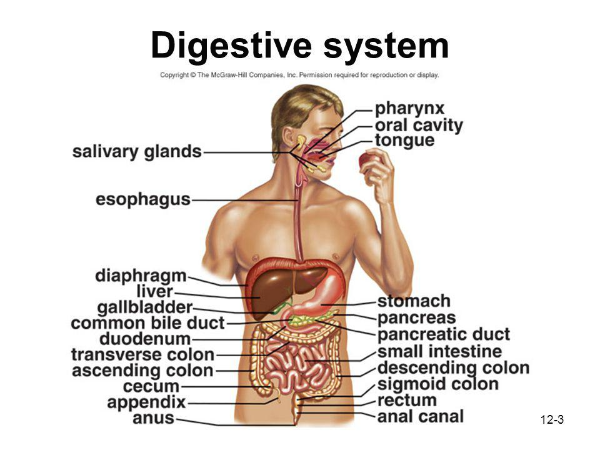 사진 출처 : SlidePlayer Digestive system 소화기 계통 Pharynx 인두 Oral cavity 구강(口腔) Tongue 혓바닥 Salivary glands 침샘, 타액선(唾液腺). Diaphragm 횡격막 Liver 간장. 간 Gallbladder 쓸개. 담낭. Common bile duct 총담관 (總膽管) 간에서 만들어진 쓸개즙과 쓸개에서 나오는 쓸개관이 합쳐서 샘창자로 주입되는 관.=온쓸개관. Duodenum 십이지장. Transverse colon 횡행 결장(橫行結腸). Ascending colon 상행 결장(上行結腸). Cecum 맹장. Appendix 부속물. 충수(蟲垂) Anus 항문 Stomach 위(胃) Pancreas 췌장(膵臟). Pancreatic duct 췌관(膵管), 췌장[이자]관 Small intestine 소장 Descending colon 내림잘룩창자, 하행결장(下行結腸), 내림주름창자, 내림창자 Sigmoid colon S 자 결장(結腸). S상결장. Rectum 직장(直腸) Anal canal 항문관(肛門管). 소화관의 종말부분. 직장(直腸)에서 원위개구부(遠位開口部)까지 뻗어 있다. ------------------------------------------------------------------------------------------------- 블로그 주인인 제가 성장 과정을 기록하기 위한 기도를 드릴 때, 침을 십이지장 소장 대장 등에 보내주는 경우가 있다고, 느낌으로 가르쳐 주십니다.
소화 시스템 목차 소화 기계 | 계획 및 위치 | 소화 과정의 단계
소화 기계의 구성 요소 | 식욕 조절 | 영양 | 학습 목표 | 연결
동물은 대부분의 경우 음식을 크고 복잡한 분자로 섭취하며, 이는 모든 세포의 몸 전체에 분포 할 수있는 더 작은 분자 (단량체)로 분해되어야합니다. 이 중요한 기능은 소화 시스템을 구성하는 일련의 특수 기관에 의해 축적됩니다. 대표적인 소화 시스템은 그림 1에 나와 있습니다.
소화 기계 | 맨 위로 단세포 유기체는 외부 환경에서 직접 영양분을 섭취 할 수 있습니다. 대부분의 세포가 외부 환경과의 직접 접촉에서 제거 된 다세포 동물은 먹이를 얻고 분해하기위한 특수 구조를 개발했습니다. 동물은 먹이와 소화의 두 가지 과정에 의존합니다.
동물은 종속 영양 생물 이며 영양분을 흡수하거나 음식을 섭취해야합니다. 대부분의 동물 인 섭취를 먹는 사람 은 입을 사용하여 음식을 섭취합니다. 촌충과 같은 흡수성 피더 는 다른 동물의 소화계에 살며 그 동물의 영양분을 체벽을 통해 직접 흡수합니다. 굴과 홍합과 같은 필터 피더 는 주변 물에서 작은 유기체와 입자를 수집합니다. 지렁이와 흰개미와 같은 기질 피더 는 그들이 뚫고 들어가는 물질 (흙이나 나무)을 먹습니다. 진딧물과 같은 액체 공급 장치 는 식물이나 동물의 몸을 뚫고 액체를 빼냅니다.
그림 1. 대표적인 동물의 소화 시스템. Purves et al., Life : The Science of Biology , 4th Edition, Sinauer Associates ( www.sinauer.com ) 및 WH Freeman ( www.whfreeman.com )의 이미지가 허가를 받아 사용되었습니다.
계획 및 위치 | 맨 위로 소화 시스템은 혈액으로 흡수 될 수있는 영양소 분자로 음식을 아래로 휴식을 기계적, 화학적 방법을 사용합니다. 혈액에 들어가면 음식 분자는 동물의 몸에있는 모든 세포로 이동합니다.
두 가지 유형의 동물 신체 계획과 소화가 발생하는 두 위치가 있습니다. 주머니와 같은 계획은 많은 무척추 동물에서 발견되며 음식물 섭취와 폐기물 배출을위한 단일 구멍이 있습니다. 인간이 속한 동물 그룹 인 척추 동물 은 음식이 한 구멍 (입)을 통해 들어가고 폐기물이 다른 구멍 (항문)을 통해 빠져 나가는 보다 효율적인 튜브 내 튜브 계획을 사용합니다.
음식의 소화가 일어나는 곳도 다양합니다. 일부 동물은 세포 내 소화를 사용하는데 , 여기서 음식은 식세포 소포 로 분비되는 소화 효소와 함께 식균 작용 에 의해 세포 로 들어갑니다 . 이러한 유형의 소화는 스펀지, coelenterates (산호, hydras 및 그 친척) 및 대부분의 원생 동물에서 발생합니다. 세포 외 소화 는 소화 시스템의 내강 (또는 개구부)에서 발생하며 영양분 분자는 혈액이나 다른 체액으로 전달됩니다. 이보다 진보 된 소화 유형은 chordates, annelids 및 갑각류에서 발생합니다.
소화 과정의 단계 | 맨 위로 대부분의 식품은 전분, 단백질 및 지방과 같은 다양한 유기 거대 분자로 구성됩니다. 이러한 분자는 개별 모노머 단위로 만들어진 폴리머입니다 (이전 장에서 논의 됨). 이러한 큰 분자를 더 작은 구성 요소로 나누는 작업은 다음과 같습니다.
운동 : 소화 시스템을 통해 음식을 추진합니다. 분비물 : 특정 자극에 반응하여 소화액 분비 소화 : 음식물을 원형질막을 통과 할 수있을 정도로 작은 분자 성분으로 분해 흡수 : 분자가 체내로 이동하고 체내로 이동 제거 : 소화되지 않은 음식 및 폐기물 제거 우리가 느슨하게 "소화"라고 부르는 동안 세 가지 과정이 발생합니다. 적절한 소화는 음식을 혈액으로 통과 할 수있을만큼 작은 입자 / 분자로 음식을 기계적 및 화학적으로 분해하는 것입니다. 흡수는 식품 단량체가 혈류로 통과하는 것입니다. 동화는 음식 분자가 체세포로 이동하는 것입니다.
소화 기계의 구성 요소 | 맨 위로 그림 2에 표시된 것처럼 인간의 소화 시스템은 입에서 항문까지 뻗어있는 코일 형 근육 튜브 (완전히 확장 된 경우 길이 6-9m)입니다. 이 길이를 따라 입, 인두, 식도, 위, 소장, 대장 및 항문과 같은 몇 가지 특수 구획이 발생합니다. 부속 소화 기관은 침샘, 췌장의 일부, 간 및 담낭 (담즙 계 ) 과 같은 일련의 관에 의해 주요 시스템에 연결됩니다 .
그림 2. 인간의 소화 시스템. Purves et al., Life : The Science of Biology , 4th Edition, Sinauer Associates ( www.sinauer.com ) 및 WH Freeman ( www.whfreeman.com )의 이미지가 허가를 받아 사용되었습니다.
입과 인두 기계적 쇠약은 씹는 (치아)과 혀의 작용에 의해 입에서 시작됩니다. 제작하여 전분의 화학적 분해 타액 아밀라제 로부터 침샘 . 이 음식물과 타액의 혼합물은 인두 와 식도 로 밀려납니다 . 식도는 근육 수축 ( 연동 운동 )이 음식을 위로 밀어내는 근육 관입니다 .
입안에서 이빨, 턱 및 혀는 그림 3에서와 같이 음식을 더 작은 입자로 기계적으로 분해하기 시작합니다. 새 (딱딱한 부리로 이빨을 잃은)를 제외한 대부분의 척추 동물은 찢어지고 갈기위한 이빨이 있습니다. 씹는 음식. 혀는 씹거나 삼키는 동안 음식을 조작합니다. 포유류는 혀에 미뢰가 밀집되어 있습니다.
침샘은 전분을 포도당으로 분해하는 효소 인 침 아밀라아제를 분비합니다. 점액은 음식을 적시고 식도를 윤활합니다. 타액의 중탄산염 이온은 식품의 산을 중화합니다.
삼키면 음식이 입에서 인두를 통해 식도로 이동 한 다음 위장으로 이동합니다.
1 단계 : 씹고 축축한 음식 덩어리 인 볼 루스가 혀에 의해 나방 뒤쪽으로 이동합니다. 인두에서 볼 루스는 음식물이 폐로 들어가는 것을 막고 볼 루스가 식도로 향하도록하는 비자발적 삼킴 반사를 유발합니다. 2 단계 : 식도의 근육은 식도를 감싸는 평활근의 비자발적 근육 수축 (페리 탈 시스)의 파동에 의해 볼 루스를 추진합니다. 연동 운동은 그림 4에 나와 있습니다. 3 단계 : 볼 루스는 위식도 괄약근을 통해 위로 전달됩니다. 속쓰림은이 괄약근을 통해 누출되는 위액에 의한 식도 자극으로 인해 발생합니다. 그림 3. 인후의 구조와 삼키는 역학. Purves et al., Life : The Science of Biology , 4th Edition, Sinauer Associates ( www.sinauer.com ) 및 WH Freeman ( www.whfreeman.com )의 허가를 받아 이미지 제공.
그림 4. 연동 운동과 입에서 위로 음식의 이동. Purves et al., Life : The Science of Biology , 4th Edition, Sinauer Associates ( www.sinauer.com ) 및 WH Freeman ( www.whfreeman.com )의 허가를 받아 이미지 제공.
위 (또는 Churn, Churn, Churn) 식사 중에 위는 50-100 밀리리터의 빈 용량에서 점차적으로 1 리터의 용량으로 채워집니다. 불편 함의 대가로 위는 2 리터 이상을 유지하기 위해 팽창 할 수 있습니다.
그림 5에서와 같이 상피 세포 는 위의 안쪽 표면에 선을 긋고 하루에 약 2 리터의 위액을 분비합니다. 위액에는 염산, 펩시노겐 및 점액이 포함되어 있습니다 . 소화에 중요한 성분. 분비물은 신경 (냄새, 생각, 카페인)과 내분비 신호에 의해 조절됩니다. 위는 염산과 펩신을 분비 합니다. 염산 (HCl)은 위의 pH를 낮추어 펩신이 활성화됩니다. 펩신은 단백질의 펩타이드로의 가수 분해를 제어하는 효소입니다. 위는 또한 음식을 기계적으로 휘젓습니다. 위의 산과 음식이 혼합 된 Chyme은 위를 떠나 소장으로 들어갑니다.
그림 5. 포유류 X2000의 위 내벽에 대한 주사 전자 현미경 사진. 이 이미지는 http://130.102.208.100/FMRes/FMPro?-db=images.fp3&key=32819&-img 에서 가져온 것으로 Nanoworld의 허가를 받아 사용되었습니다.
염산은 소화에서 직접 기능하지 않습니다. 미생물을 죽이고 위 pH를 1.5에서 2.5 사이로 낮 춥니 다. 펩시노겐을 활성화합니다. 펩시노겐은 단백질 소화를 시작하는 효소입니다. 펩시노겐은 위 구덩이에 있는 세포에서 생성됩니다 . 그것은 분자의 일부를 절단하여 활성화되어 위장에서 소화되는 동안 단백질 분자에서 펩티드 조각을 분리하는 효소 펩신을 생성합니다.
입안의 타액 아밀라아제에 의해 시작된 탄수화물 소화는 위로 전달 될 때 볼 루스에서 계속됩니다. 볼 루스는 위의 3 분의 1에서 산성 카임으로 분해되어 위의 산도가 추가로 탄수화물 분해를 억제 할 수 있도록합니다. 펩신에 의한 단백질 소화가 시작됩니다.
알코올과 아스피린은 위벽을 통해 혈액으로 흡수됩니다.
상피 세포는 세포와 위산 사이에 보호 장벽을 형성하는 점액을 분비합니다. 펩신은 점액과 접촉하면 비활성화됩니다. 중탄산염 이온은 위를 감싸는 세포 근처의 산도를 감소시킵니다. 단단한 접합부 는 상피 위 내벽 세포를 함께 연결하여 위산이 통과하는 것을 추가로 줄이거 나 방지합니다.
궤양 이러한 보호 메커니즘이 실패하면 소화성 궤양 이 발생합니다. 출혈성 궤양은 조직 손상이 너무 심해서 위장으로 출혈이 발생할 때 발생합니다. 천공성 궤양은 위벽에 구멍이 생기는 생명을 위협하는 상황입니다. 모든 소화성 궤양의 최소 90 %는 헬리코박터 파일로리 에 의해 발생합니다 . 스트레스와 아스피린을 포함한 다른 요인들도 궤양을 유발할 수 있습니다.
소장 소장 최종 소화 흡수가 발생하는 위치를도 6에 도시 된이있다. 소장은 길이가 3 미터가 넘는 코일 튜브입니다. 코일과 폴딩 플러스 융모는이 3m 튜브에 500-600m 길이 튜브의 표면적을 제공합니다. 단백질과 탄수화물의 최종 소화가 이루어져야하며 지방은 아직 소화되지 않았습니다. 융모 에는 펩티드와 당의 소화를 완료하는 장 효소를 생성하는 세포가 있습니다. 흡수 과정은 소장에서도 발생합니다. 음식은 소장으로 들어갈만큼 작은 입자로 분해되었습니다. 설탕과 아미노산은 각 융모의 모세 혈관을 통해 혈류로 들어갑니다. 글리세롤과 지방산은 림프계로 들어갑니다. 흡수는 활성 수송이며 세포 에너지를 필요로합니다.
그림 6. 소장의 구조 및 세부 사항. Purves et al., Life : The Science of Biology , 4th Edition, Sinauer Associates ( www.sinauer.com ) 및 WH Freeman ( www.whfreeman.com )의 이미지가 허가를 받아 사용되었습니다.
음식은 유문 괄약근에 대해 산성-엽 혼합물을 추진하는 연동 파에 의해 위의 아래 부분에서 혼합됩니다. 위의 수축이 증가하면 위가 1 ~ 2 시간 동안 배출됨에 따라 음식이 괄약근을 통해 소장으로 밀려납니다. 고지방 식단은이 기간을 상당히 증가시킵니다.
소장은 영양소의 소화 및 흡수를위한 주요 부위입니다. 소장은 길이가 최대 6 미터이고 너비가 2-3 센티미터입니다. 윗부분 인 십이지장 은 소화에서 가장 활동적입니다. 간과 췌장의 분비물은 십이지장의 소화에 사용됩니다. 십이지장의 상피 세포는 수분 점액을 분비합니다. 췌장은 소화 효소와 위산을 중화시키는 중탄산염을 분비합니다. 간은 담즙을 생성하며, 담즙은 담관으로 들어가 십이지장으로 들어가기 전에 담낭에 저장됩니다.
탄수화물, 단백질 및 지방의 소화는 소장에서 계속됩니다. 전분과 글리코겐은 소장 효소에 의해 말토오스로 분해됩니다. 프로테아제는 췌장에서 분비되는 효소로 단백질을 작은 펩타이드 조각과 아미노산으로 계속 분해합니다.
담즙은 지방을 유화하여 리파아제 에 의해 작용할 수있을 때까지 점진적으로 더 작은 지방 구로 분해를 촉진합니다 . 담즙에는 콜레스테롤, 인지질, 빌리루빈 및 혼합 염이 포함되어 있습니다. 지방은 탄수화물과 단백질과 달리 소장에서 완전히 소화됩니다.
대부분의 흡수는 십이지장과 제 즈음 (소장의 2/3) 에서 발생합니다 . 장의 내부 표면에는 흡수를위한 표면적의 3 배 이상인 원형 주름이 있습니다. 상피 세포로 덮인 융모 는 표면적 을 10 배 더 증가시킵니다 . 상피 세포는 표면적을 더욱 증가시키는 미세 융모 가 늘어서 있습니다. 6 미터 길이의 튜브는 표면적이 300 평방 미터입니다.
각 융모에는 브러시 테두리 로 알려진 상피 세포 위에 형성되는 미세 융모로 덮인 소장 개구부 내부에 인접한 표면이 있습니다 . 각 융모에는 작은 세동맥에 의해 공급되는 모세관 네트워크가 있습니다. 흡수 된 물질은 일반적으로 수동 수송에 의해 브러시 경계를 통해 모세관으로 전달됩니다.
맥아당, 자당 및 유당은 소장에 존재하는 주요 탄수화물입니다. 그들은 미세 융모에 흡수됩니다. 전분은 다른 곳에서 2 개의 포도당 단위 (말토오스)로 분해됩니다. 세포의 효소는이 이당류 를 단당류 로 변환 한 다음 세포 를 떠나 모세관으로 들어갑니다. 유당 불내증 은 장 세포에서 생산되는 효소 락타아제의 유전 적 결여로 인해 발생합니다.
펩티드 조각과 아미노산은 활성 수송에 의해 상피 세포막을 통과합니다 . 세포 내부에서 그들은 아미노산으로 분해되어 모세관으로 들어갑니다. 글루텐 장 병증은 밀에서 발견되는 단백질 인 글루텐을 흡수하지 못하는 것입니다.
소화 된 지방은 잘 녹지 않습니다. 담즙 염은 지방을 둘러싸고 그림 7과 같이 상피 세포로 들어갈 수있는 미셀 을 형성 합니다. 담즙 염은 루멘으로 돌아가 과정을 반복합니다. 지방 소화는 보통 음식이 소장 의 회장 (하위 1/3)에 도달 할 때 완료됩니다 . 담즙 염은 회장에 흡수되어 간과 담낭에서 재활용됩니다. 지방은 상피 세포에서 융모를 통과하는 작은 림프관으로 전달됩니다.
그림 7. 소장의 세포에 의한 지질 흡수. Purves et al., Life : The Science of Biology , 4th Edition, Sinauer Associates ( www.sinauer.com ) 및 WH Freeman ( www.whfreeman.com )의 이미지가 허가를 받아 사용되었습니다.
간 및 담낭 간은 그림 8과 같이 담즙을 생성하여 간관을 통해 소장으로 보냅니다. 담즙에는 지방을 유화시켜 효소 분해에 취약한 담즙 염이 포함되어 있습니다. 소화 기능 외에도 간은 몇 가지 다른 역할을합니다. 1) 혈액 해독; 2) 혈액 단백질 합성; 3) 오래된 적혈구의 파괴 및 헤모글로빈 을 담즙 성분으로 전환 ; 4) 담즙 생성; 5) 포도당을 글리코겐 으로 저장하고 혈당 수치가 떨어지면 방출됩니다. 및 6) 아미노기 및 암모니아로부터 요소 생산.
그림 8. 간 및 관련 기관 및 소화 시스템과의 연결. Purves et al., Life : The Science of Biology , 4th Edition, Sinauer Associates ( www.sinauer.com ) 및 WH Freeman ( www.whfreeman.com )의 이미지가 허가를 받아 사용되었습니다.
담낭은 나중에 방출하기 위해 과도한 담즙을 저장합니다. 우리는 담낭 없이도 살 수 있습니다. 사실 많은 사람들이 담낭을 제거했습니다. 그러나 결점은 담낭에 저장된 담즙을 더 이상 사용할 수 없기 때문에 그들이 먹는 음식에있는 지방의 양을 인식 할 필요가 있다는 것입니다.
글리코겐 은 그림 9와 같이 포도당 분자 사슬로 구성된 다당류입니다. 식물에서 전분은 포도당의 저장 형태 인 반면 동물은 동일한 목적으로 글리코겐을 사용합니다. 혈중 포도당 수치가 낮 으면 간으로 이동하여 글리코겐이 포도당으로 분해되는 것을 자극하는 글루카곤 과 같은 호르몬이 분비 되어 혈당 수치가 높아집니다. 포도당이나 글리코겐을 사용할 수없는 경우 아미노산은 간에서 포도당으로 전환됩니다. 탈 아미 노화 과정은 아미노산에서 아미노기를 제거합니다. 우레아가 형성되고 혈액을 통해 신장으로 전달되어 신체에서 배출됩니다. 반대로 인슐린 호르몬 은 글루 소스가 간 세포로 흡수되고 글리코겐으로 형성되는 것을 촉진합니다.
그림 9. 글리코겐 구조. 글리코겐을 형성하기 위해 연결된 개별 포도당 분자에 유의하십시오. http://www.bio.brandeis.edu/classes/bio18/glycogen.gif의 이미지 .
간 질환 황달은 피부의 특징적인 노란색 색조가 혈중 헤모글로빈 분해 산물 과다로 인해 발생하며, 이는 간이 제대로 기능하지 않는다는 신호입니다. 황달은 담관 막힘 및 간염으로 인한 손상으로 인해 간 기능이 손상되었을 때 발생할 수 있습니다 .
A 형, B 형, C 형 간염은 모두 간 손상을 일으킬 수있는 바이러스 성 질환입니다. 다른 바이러스 질환과 마찬가지로 주요 치료 노력은 바이러스 원인 제거가 아닌 증상 치료에 중점을 둡니다. A 형 간염은 일반적으로 갑작스런 발열, 불쾌감, 메스꺼움, 식욕 부진 및 복부 불편 함으로 표시되는 경미한 질병입니다. 황달은 며칠 동안 후속 조치를 취합니다. A 형 간염을 일으키는 바이러스는 오염 된 음식과 물도 전염을 촉진 할 수 있지만 배설물 오염에 의해 일차적으로 전염됩니다. 미국에서 드문 질병 인 B 형 간염은 수억 명의 사람들이 감염 될 가능성이있는 아시아 일부 지역에서 발병합니다.
B 형 간염은 성적 접촉뿐만 아니라 혈액 및 혈액 제제에 의해 전염 될 수 있습니다. 선진국의 혈액 공급은 수년 동안이 질병을 일으키는 바이러스에 대해 선별되었으며 수혈을 통한 전염은 드뭅니다. HBV 감염의 위험은 이종성으로도 전염되지만 난잡한 동성애 남성들 사이에서 높습니다. 콘돔의 올바른 사용은 전파의 위험을 줄이거 나 없애는 것으로 생각됩니다. B 형 간염 감염 예방을위한 효과적인 백신이 있습니다. 만성 B 형 간염이있는 일부 개인은 간경변증이 발생할 수 있습니다. 만성 B 형 간염 환자는 원발성 간암에 걸릴 위험이 높습니다. 이러한 유형의 암은 미국에서 비교적 드물지만 세계에서 암 사망의 주요 원인입니다.
C 형 간염은 전 세계적으로 약 1 억 7 천만 명, 미국에서는 4 백만 명에게 영향을 미칩니다. 바이러스는 주로 혈액 및 혈액 제품에 의해 전염됩니다. 대부분의 감염된 사람들은 1990 년 이전 (C 형 간염 바이러스에 대한 혈액 공급 검사가 시작되었을 때) 이전에 수혈을 받았거나 정맥 주사 약물을 사용했습니다. 일부일처 제 부부 사이에서 성적인 전파가 발생할 수 있지만 (희귀), 난잡한 사람들에게는 감염이 훨씬 더 흔합니다. 드물게 C 형 간염은 급성 질환과 심지어 간부전을 유발합니다. 간경변증이있는 C 형 간염 환자의 약 20 %는 또한 심각한 간 질환을 앓게됩니다. C 형 간염으로 인한 간경변은 현재 미국에서 간 이식이 필요한 주요 원인입니다. C 형 간염으로 인한 간경변증이있는 사람은 또한 원발성 간암에 걸릴 가능성이 높습니다. C 형 간염에 대한 현재의 모든 치료는 강력한 항 바이러스 인터페론 알파의 다양한 제제를 사용합니다. 그러나 질병에 걸린 모든 환자가 치료에 적합한 것은 아니므로 감염된 개인은 정기적으로 의사와 상담해야합니다.
간경변증은 일반적으로 알코올 중독자에게서 발생하며, 알코올 중독자는 분해 될 알코올의 양으로 인해 간을 스트레스 상황에 놓이게됩니다. 간경변은 간이 생화학 적 기능을 수행하지 못하게 할 수 있습니다. 혈액 응고를 담당하는 화학 물질은 혈액의 주요 단백질 인 알부민과 마찬가지로 간에서 합성됩니다. 간은 또한 담즙 성분을 만들거나 수정합니다. 순환계의 혈액은 간을 통과하므로 콜레스테롤 대사와 단백질과 지방을 포도당으로 전환하는 등 신체의 많은 대사 기능이 주로 그곳에서 발생합니다. 간경변은 독소, 염증 및 기타 원인으로 인한 간세포 손상으로 인한 질병입니다. 간 세포는 비정상적인 패턴으로 재생되어 주로 섬유 조직으로 둘러싸인 결절을 형성합니다. 간 구조의 변화는 혈류를 감소시켜 이차적 인 합병증을 유발할 수 있습니다. 간경변에는 알코올성 간 질환, 심각한 형태의 일부 바이러스 성 간염, 울혈 성 심부전, 기생충 감염 (예 : 주혈 흡충증), 독소 나 약물에 대한 장기 노출 등 많은 원인이 있습니다.
췌장 췌장 췌관 통해 작은 intestive 때문에, 유미 즙을 중화 췌액을 보낸다. 이 소화 기능 외에도 췌장은 글루카곤 및 인슐린과 같은 여러 호르몬의 생산 부위입니다.
췌장은 소화 효소를 소장과 내분비 세포 클러스터 ( 췌도 ) 로 분비하는 외분비 세포를 포함 합니다. 이 섬은 혈당 수치를 조절 하는 호르몬 인슐린 과 글루카곤을 분비합니다 .
식사 후 혈당 수치가 상승하여 인슐린이 분비되어 세포가 포도당을 흡수하고 간 및 골격근 세포가 탄수화물 글리코겐 을 형성합니다 . 혈중 포도당 수치가 떨어지면 추가 인슐린 생산이 억제됩니다. 글루카곤은 글리코겐을 포도당으로 분해하고, 다시 혈당으로 방출되어 항상성 범위 내에서 포도당 수준을 유지합니다. 글루카곤 생산은 혈당 수치가 떨어지면 자극되고 증가하면 억제됩니다.
당뇨병 부적절한 수준의 인슐린으로 인해 발생합니다. 제 1 형 당뇨병은 종종 유전 적 원인으로 인해 부적절한 수준의 인슐린 분비가 특징입니다. 유형 II는 일반적으로 유전 적 원인과 환경 적 원인 모두에서 성인에서 발생합니다. 인슐린 부족보다는 인슐린에 대한 표적의 반응 상실이 이러한 유형의 당뇨병을 유발합니다. 당뇨병은 눈, 순환계, 신경계 및 신장 기능 장애를 일으킬 수 있습니다. 당뇨병은 미국에서 실명의 두 번째 주요 원인입니다. 치료에는 매일 인슐린 주사, 메트포르민과 같은 경구 약물 투여, 혈당 수치 모니터링 및 조절식이 포함될 수 있습니다. 제 1 형 당뇨병은 유전자 치료 / 줄기 세포 연구의 발전으로 언젠가는 치료 될 수 있습니다. 최근에인지 된 상태는 당뇨병 전증으로 알려져 있습니다. 신체가 점차 인슐린에 대한 민감성을 잃어 결국 제 2 형 당뇨병으로 이어집니다. Ora; 약물,식이 요법 및 행동 (즉, 운동 !!!) 변화는 당뇨 발병을 완전히 연기하지 않으면 지연되는 것으로 생각됩니다.
미국에서 암 사망의 다섯 번째 주요 원인은 거의 항상 치명적인 췌장암입니다. 과학자들은 매년 25,000 명이이 질병으로 사망 할 수 있다고 추정합니다. 표준 치료법은 효과가 없지만 일부 유망한 길은 암세포의 유전체학 및 분자 생물학의 발전으로 열릴 수 있습니다.
대장 대장은 결장, 맹장,로 구성되어 부록 및 직장. 대장의 물질은 대부분 소화가 안되는 잔류 물과 액체입니다. 움직임은 내용물을 앞뒤로 섞는 비자발적 수축과 대장을 통해 물질을 이동시키는 추진 적 수축으로 인해 발생합니다. 대장은 척추 동물에서 세 가지 기본 기능을 수행합니다. 1) 소화 된 음식에서 물과 전해질을 회수합니다. 2) 대변의 형성 및 저장; 3) 미생물 발효 : 대장은 놀라운 미생물 군집을 지원합니다. 이러한 미생물은 척추 동물이 소화 할 수없는 많은 분자를 소화 할 수있는 효소를 생산합니다.
대장의 분비물은 상피 조직을 보호하고 박테리아 대사에 의해 생성 된 산을 중화시키는 알칼리성 점액입니다. 물, 염분 및 비타민이 흡수되고 루멘의 나머지 내용물은 대변을 형성 합니다 (주로 셀룰로오스, 박테리아, 빌리루빈). 대장균 과 같은 대장의 박테리아 는 흡수되는 비타민 (비타민 K 포함)을 생성 합니다.
식욕 조절 | 맨 위로 뇌 의 시상 하부 에는 배고픔을 조절하는 두 개의 센터가 있습니다. 하나는 식욕 센터이고 다른 하나는 포만 센터입니다.
가스트린 , 세크레틴 , 콜레시스토키닌 은 다양한 소화 단계를 조절하는 호르몬입니다. 위장에 단백질이 존재하면 가스트린 분비가 촉진되어 위산 분비가 증가하고 소화관의 이동성이 음식물을 이동하게됩니다. 음식물이 십이지장으로 전달되면 세크레틴이 생성되고, 이는 차례로 췌장에서 알칼리성 분비물의 방출을 촉진하고 산이 중화 될 때까지 음식물이 장으로 더 이상 통과하지 못하도록합니다. 콜레시스토키닌 (CCK)은 지방에 반응하여 장 상피에서 방출되며 담낭에서 담즙이 방출되고 췌장에서 리파아제 (지방 소화 효소)가 방출됩니다.
영양 | 맨 위로 영양은 음식의 구성, 에너지 함량 및 천천히 (또는 전혀) 합성 된 유기 분자를 다룹니다. Chemotrophs 는 무기 화학 반응에서 에너지를 얻는 유기체 (대부분 박테리아)입니다. Phototrophs 는 햇빛 에너지를 설탕이나 다른 유기 분자로 변환합니다. Heterotrophs는 음식에서 유기 분자가 분해되어 에너지를 얻기 위해 먹습니다.
다량 영양소 는 매일 대규모로 필요한 식품입니다. 여기에는 탄수화물, 지질 및 아미노산이 포함됩니다. 물은 필수이며 올바른 물 균형은 신체의 적절한 기능을 위해 필수입니다.
식단의 약 60 %는 우유, 육류, 채소, 곡물 및 곡물 제품과 같은 식품에서 얻은 탄수화물이어야합니다. 식단에는 매일 최소 100g의 탄수화물이 포함되어야합니다. 그러나 최근에는 탄수화물의 양을 줄 이도록 제안하는 새로운 권장 사항이 개발되었습니다. 이 주제에 대한 자세한 프레젠테이션은 http://health.discovery.com/diseasesandcond/encyclopedia/2935.html 에서 찾을 수 있습니다 .
단백질은 아미노산으로 구성된 폴리머입니다. 단백질은 육류, 우유, 가금류, 생선, 시리얼 곡물 및 콩에서 발견됩니다. 그들은 세포 성장과 복구에 필요합니다. 단백질에는 20 개의 아미노산이 있으며, 그중 인간은 11 개를 만들 수 있습니다. 나머지 9 개는 식단에 반드시 공급되어야하는 필수 아미노산입니다. 일반적으로 단백질은 에너지로 사용되지 않지만 굶주림 (또는 저탄수화물 다이어트) 중에는 근육 단백질이 에너지로 분해됩니다. 과도한 단백질은 에너지로 사용되거나 지방으로 전환 될 수 있습니다.
지질과 지방은 가장 큰 에너지 생산량을 생성하므로 많은 수의 식물과 동물이 과도한 음식 에너지를 지방으로 저장합니다. 지방과 지방은 기름, 육류, 버터 및 식물 (예 : 아보카도 및 땅콩)에 존재합니다. 리놀레산과 같은 일부 지방산은 필수이며 식단에 포함되어야합니다. 장에 존재할 때 지질은 비타민 A, D, E 및 K의 섭취를 촉진합니다.
비타민은 대사 반응에 필요한 유기 분자입니다. 그들은 일반적으로 신체에서 만들 수 없으며 미량으로 필요합니다. 비타민은 효소 보조 인자 또는 보조 효소로 작용할 수 있습니다 . 일부 비타민은 지방에 용해되고 일부는 물에 용해됩니다.
미네랄 은 세포와 조직의 구성 요소로서 정상적인 신진 대사, 신경 전도 및 근육 수축에 필요한 미량 원소입니다. 그들은 식단에서만 얻을 수 있습니다. 철 (헤모글로빈), 요오드 (티록신), 칼슘 (뼈) 및 나트륨 (신경 메시지 전달)이 미네랄의 예입니다.
영양소와 건강 사이에는 양적 관계가 있습니다. 불균형은 질병을 유발할 수 있습니다. 많은 연구에서 영양이 심혈관 질환, 고혈압 및 암의 주요 요인이라는 결론을 내 렸습니다.
THE DIGESTIVE SYSTEM Table of Contents Digestive System | Plans and Locations | Stages in the Digestive Process
Components of the Digestive System | Regulation of Appetite | Nutrition | Learning Objectives | Links
Animals, for the most part, ingest their food as large, complex molecules that must be broken down into smaller molecules (monomers) that can then be distributed throughout the body of every cell. This vital function is accpomplished by a series of specialized organs that comprise the digestive system. Representative digestive systems are shown in Figure 1.
Digestive System | Back to Top Single-celled organisms can directly take in nutrients from their outside environment. Multicellular animals, with most of their cells removed from contact directly with the outside environment, have developed specialized structures for obtaining and breaking down their food. Animals depend on two processes: feeding and digestion.
Animals are heterotrophs, they must absorb nutrients or ingest food sources. Ingestive eaters, the majority of animals, use a mouth to ingest food. Absorptive feeders, such as tapeworms, live in a digestive system of another animal and absorb nutrients from that animal directly through their body wall. Filter feeders, such as oysters and mussels, collect small organisms and particles from the surrounding water. Substrate feeders, such as earthworms and termites, eat the material (dirt or wood) they burrow through. Fluid feeders, such as aphids, pierce the body of a plant or animal and withdraw fluids.
Figure 1. The digestive systems of representative animals. Images from Purves et al., Life: The Science of Biology, 4th Edition, by Sinauer Associates (www.sinauer.com) and WH Freeman (www.whfreeman.com), used with permission.
Plans and Locations | Back to Top The digestive system uses mechanical and chemical methods to break food down into nutrient molecules that can be absorbed into the blood. Once in the blood, the food molecules are routed to every cell in the animal's body.
There are two types of animal body plans as well as two locations fordigestion to occur. Sac-like plans are found in many invertebrates, who have a single opening for food intake and the discharge of wastes. Vertebrates, the animal group humans belong to, use the more efficient tube-within-a-tube plan with food entering through one opening (the mouth) and wastes leaving through another (the anus).
Where the digestion of the food happens is also variable. Some animals use intracellular digestion, where food is taken into cells by phagocytosis with digestive enzymes being secreted into the phagocytic vesicles. This type of digestion occurs in sponges, coelenterates (corals, hydras and their relatives) and most protozoans. Extracellular digestion occurs in the lumen (or opening) of a digestive system, with the nutrient molecules being transferred to the blood or some other body fluid. This more advanced type of digestion occurs in chordates, annelids, and crustaceans.
Stages in the Digestive Process | Back to Top Food for the most part consists of various organic macromolecules such as starch, proteins, and fats. These molecules are polymers made of individual monomer units (as discussed in an earlier chapter). Breaking these large molecules into smaller components involves:
movement: propels food through the digestive system secretion: release of digestive juices in response to a specific stimulus digestion: breakdown of food into molecular components small enough to cross the plasma membrane absorption: passage of the molecules into the body's interior and their passage throughout the body elimination: removal of undigested food and wastes Three processes occur during what we loosely refer to as "digestion". Digestion proper, which is the mechanical and chemical breakdown of food into particles/molecules small enough to pass into the blood. Absorption is the passage of food monomers into the blood stream. Assimilation is the passage of the food molecules into body cells.
Components of the Digestive System | Back to Top The human digestive system, as shown in Figure 2, is a coiled, muscular tube (6-9 meters long when fully extended) stretching from the mouth to the anus. Several specialized compartments occur along this length: mouth, pharynx, esophagus, stomach, small intestine, large intestine, and anus. Accessory digestive organs are connected to the main system by a series of ducts: salivary glands, parts of the pancreas, and the liver and gall bladder (bilary system).
Figure 2. The human digestive system. Images from Purves et al., Life: The Science of Biology, 4th Edition, by Sinauer Associates (www.sinauer.com) and WH Freeman (www.whfreeman.com), used with permission.
The Mouth and Pharynx Mechanical breakdown begins in the mouth by chewing (teeth) and actions of the tongue. Chemical breakdown of starch by production of salivary amylase from the salivary glands. This mixture of food and saliva is then pushed into the pharynx and esophagus. The esophagus is a muscular tube whose muscular contractions (peristalsis) propel food to the stomach.
In the mouth, teeth, jaws and the tongue begin the mechanical breakdown of food into smaller particles, as shown in Figure 3. Most vertebrates, except birds (who have lost their teeth to a hardened bill), have teeth for tearing, grinding and chewing food. The tongue manipulates food during chewing and swallowing; mammals have tastebuds clustered on their tongues.
Salivary glands secrete salivary amylase, an enzyme that begins the breakdown of starch into glucose. Mucus moistens food and lubricates the esophagus. Bicarbonate ions in saliva neutralize the acids in foods.
Swallowing moves food from the mouth through the pharynx into the esophagus and then to the stomach.
Step 1: A mass of chewed, moistened food, a bolus, is moved to the back of the moth by the tongue. In the pharynx, the bolus triggers an involuntary swallowing reflex that prevents food from entering the lungs, and directs the bolus into the esophagus. Step 2: Muscles in the esophagus propel the bolus by waves of involuntary muscular contractions (peristalsis) of smooth muscle lining the esophagus. Peristalsis is shown in Figure 4. Step 3: The bolus passes through the gastroesophogeal sphincter, into the stomach. Heartburn results from irritation of the esophagus by gastric juices that leak through this sphincter. Figure 3. Structure of the throat and the mechanics of swallowing. Image from Purves et al., Life: The Science of Biology, 4th Edition, by Sinauer Associates (www.sinauer.com) and WH Freeman (www.whfreeman.com), used with permission.
Figure 4. Peristalsis and the movement of food from the mouth to the stomach. Image from Purves et al., Life: The Science of Biology, 4th Edition, by Sinauer Associates (www.sinauer.com) and WH Freeman (www.whfreeman.com), used with permission.
The Stomach (or Churn, Churn, Churn) During a meal, the stomach gradually fills to a capacity of 1 liter, from an empty capacity of 50-100 milliliters. At a price of discomfort, the stomach can distend to hold 2 liters or more.
Epithelial cells line inner surface of the stomach, as shown in Figure 5, and secrete about 2 liters of gastric juices per day. Gastric juice contains hydrochloric acid, pepsinogen, and mucus; ingredients important in digestion. Secretions are controlled by nervous (smells, thoughts, and caffeine) and endocrine signals. The stomach secretes hydrochloric acid and pepsin. Hydrochloric acid (HCl) lowers pH of the stomach so pepsin is activated. Pepsin is an enzyme that controls the hydrolysis of proteins into peptides. The stomach also mechanically churns the food. Chyme, the mix of acid and food in the stomach, leaves the stomach and enters the small intestine.
Figure 5. Scanning electron micrograph of the stomach lining of a mammal, X2000. This image is from http://130.102.208.100/FMRes/FMPro?-db=images.fp3&key=32819&-img, used by permission of Nanoworld.
Hydrochloric acid does not directly function in digestion: it kills microorganisms, lowers the stomach pH to between 1.5 and 2.5; and activates pepsinogen. Pepsinogen is an enzyme that starts protein digestion. Pepsinogen is produced in cells that line the gastric pits. It is activated by cleaving off a portion of the molecule, producing the enzyme pepsin that splits off fragments of peptides from a protein molecule during digestion in the stomach.
Carbohydrate digestion, begun by salivary amylase in the mouth, continues in the bolus as it passes to the stomach. The bolus is broken down into acid chyme in the lower third of the stomach, allowing the stomach's acidity to inhibit further carbohydrate breakdown. Protein digestion by pepsin begins.
Alcohol and aspirin are absorbed through the stomach lining into the blood.
Epithelial cells secrete mucus that forms a protective barrier between the cells and the stomach acids. Pepsin is inactivated when it comes into contact with the mucus. Bicarbonate ions reduce acidity near the cells lining the stomach. Tight junctions link the epithelial stomach-lining cells together, further reducing or preventing stomach acids from passing.
Ulcers Peptic ulcers result when these protective mechanisms fail. Bleeding ulcers result when tissue damage is so severe that bleeding occurs into the stomach. Perforated ulcers are life-threatening situations where a hole has formed in the stomach wall. At least 90% of all peptic ulcers are caused by Helicobacter pylori. Other factors, including stress and aspirin, can also produce ulcers.
The Small Intestine The small intestine, shown in Figure 6, is where final digestion and absorption occur. The small intestine is a coiled tube over 3 meters long. Coils and folding plus villi give this 3m tube the surface area of a 500-600m long tube. Final digestion of proteins and carbohydrates must occur, and fats have not yet been digested. Villi have cells that produce intestinal enzymes which complete the digestion of peptides and sugars. The absorption process also occurs in the small intestine. Food has been broken down into particles small enough to pass into the small intestine. Sugars and amino acids go into the bloodstream via capillaries in each villus. Glycerol and fatty acids go into the lymphatic system. Absorption is an active transport, requiring cellular energy.
Figure 6. Structure and details of the small intestine. Images from Purves et al., Life: The Science of Biology, 4th Edition, by Sinauer Associates (www.sinauer.com) and WH Freeman (www.whfreeman.com), used with permission.
Food is mixed in the lower part of the stomach by peristaltic waves that also propel the acid-chyme mixture against the pyloric sphincter. Increased contractions of the stomach push the food through the sphincter and into the small intestine as the stomach eempties over a 1 to 2 hour period. High fat diets significantly increase this time period.
The small intestine is the major site for digestion and absorption of nutrients. The small intestine is up to 6 meters long and is 2-3 centimeters wide. The upper part, the duodenum, is the most active in digestion. Secretions from the liver and pancreas are used for digestion in the duodenum. Epithelial cells of the duodenum secrete a watery mucus. The pancreas secretes digestive enzymes and stomach acid-neutralizing bicarbonate. The liver produces bile, which is stored in the gall bladder before entering the bile duct into the duodenum.
Digestion of carbohydrates, proteins, and fats continues in the small intestine. Starch and glycogen are broken down into maltose by small intestine enzymes. Proteases are enzymes secreted by the pancreas that continue the breakdown of protein into small peptide fragments and amino acids.
Bile emulsifies fats, facilitating their breakdown into progressively smaller fat globules until they can be acted upon by lipases. Bile contains cholesterol, phospholipids, bilirubin, and a mix of salts. Fats are completely digested in the small intestine, unlike carbohydrates and proteins.
Most absorption occurs in the duodenum and jejeunum (second third of the small intestine). The inner surface of the intestine has circular folds that more than triple the surface area for absorption. Villi covered with epithelial cells increase the surface area by another factor of 10. The epithelial cells are lined with microvilli that further increase the surface area; a 6 meter long tube has a surface area of 300 square meters.
Each villus has a surface that is adjacent to the inside of the small intestinal opening covered in microvilli that form on top of an epithelial cell known as a brush border. Each villus has a capillary network supplied by a small arteriole. Absorbed substances pass through the brush border into the capillary, usually by passive transport.
Maltose, sucrose, and lactose are the main carbohydrates present in the small intestine; they are absorbed by the microvilli. Starch is broken down into two-glucose units (maltose) elsewhere. Enzymes in the cells convert these disaccharides into monosaccharides that then leave the cell and enter the capillary. Lactose intolerance results from the genetic lack of the enzyme lactase produced by the intestinal cells.
Peptide fragments and amino acids cross the epithelial cell membranes by active transport. Inside the cell they are broken into amino acids that then enter the capillary. Gluten enteropathy is the inability to absorb gluten, a protein found in wheat.
Digested fats are not very soluble. Bile salts surround fats to form micelles, as shown in Figure 7, that can pass into the epithelial cells. The bile salts return to the lumen to repeat the process. Fat digestion is usually completed by the time the food reaches the ileum (lower third) of the small intestine. Bile salts are in turn absorbed in the ileum and are recycled by the liver and gall bladder. Fats pass from the epithelial cells to the small lymph vessel that also runs through the villus.
Figure 7. Absorption of lipids by cells in the small intestine. Images from Purves et al., Life: The Science of Biology, 4th Edition, by Sinauer Associates (www.sinauer.com) and WH Freeman (www.whfreeman.com), used with permission.
The Liver and Gall Bladder The liver produces and sends bile to the small intestine via the hepatic duct, as illustrated in Figure 8. Bile contains bile salts, which emulsify fats, making them susceptible to enzymatic breakdown. In addition to digestive functions, the liver plays several other roles: 1) detoxification of blood; 2) synthesis of blood proteins; 3) destruction of old erythrocytes and conversion of hemoglobin into a component of bile; 4) production of bile; 5) storage of glucose as glycogen, and its release when blood sugar levels drop; and 6) production of urea from amino groups and ammonia.
Figure 8. The liver and associated organs and their connections to the digestive system. Images from Purves et al., Life: The Science of Biology, 4th Edition, by Sinauer Associates (www.sinauer.com) and WH Freeman (www.whfreeman.com), used with permission.
The gall bladder stores excess bile for release at a later time. We can live without our gall bladders, in fact many people have had theirs removed. The drawback, however, is a need to be aware of the amount of fats in the food they eat since the stored bile of the gall bladder is no longer available.
Glycogen is a polysaccharide made of chains of glucose molecules, as shown in Figure 9. In plants starch is the storage form of glucose, while animals use glycogen for the same purpose. Low glucose levels in the blood cause the release of hormones, such as glucagon, that travel to the liver and stimulate the breakdown of glycogen into glucose, which is then released into the blood(raising blood glucose levels). When no glucose or glycogen is available, amino acids are converted into glucose in the liver. The process of deamination removes the amino groups from amino acids. Urea is formed and passed through the blood to the kidney for export from the body. Conversely, the hormone insulin promotes the take-up of glusose into liver cells and its formation into glycogen.
Figure 9. Glycogen structure. Note the individual glucose molecules that are linked to form glycogen. Image from http://www.bio.brandeis.edu/classes/bio18/glycogen.gif.
Liver diseases Jaundice occurs when the characteristic yellow tint to the skin is caused by excess hemoglobin breakdown products in the blood, a sign that the liver is not properly functioning. Jaundice may occur when liver function has been impaired by obstruction of the bile duct and by damage caused by hepatitis.
Hepatitis A, B, and C are all viral diseases that can cause liver damage. Like any viral disease, the major treatment efforts focus on treatment of symptoms, not removal of the viral cause. Hepatitis A is usually mild malady indicated by a sudden fever, malaise, nausea, anorexia, and abdominal discomfort. Jaundice follows up for several days. The virus causing Hepatitis A is primarilly transmitted by fecal contamination, although contaminated food and water also can promote transmission. A rare disease in the United States, hepatitis B is endemic in parts of Asia where hundreds of millions of individuals are possibly infected.
Hepatitis B may be transmitted by blood and blood products as well as sexual contact. The blood supply in developed countries has been screened for the virus that causes this disease for many years and transmission by blood transfusion is rare. The risk of HBV infection is high among promiscuous homosexual men although it is also transmitted hetereosexually. Correct use of condoms is thought to reduce or eliminate the risk of transmission. Effective vaccines are available for the prevention of Hepatitis B infection. Some individuals with chronic hepatitis B may develop cirrhosis of the liver. Individuals with chronic hepatitis B are at an increased risk of developing primary liver cancer. Although this type of cancer is relatively rare in the United States, it is the leading cause of cancer death in the world, primarily because the virus causing it is endemic in eastern Asia.
Hepatitis C affects approximately 170 million people worldwide and 4 million in the United States. The virus is transmitted primarily by blood and blood products. Most infected individuals have either received blood transfusions prior to 1990 (when screening of the blood supply for the Hepatitis C virus began) or have used intravenous drugs. Sexual transmission can occur between monogamous couples (rare) but infection is far more common in those who are promiscuous. In rare cases, Hepatitis C causes acute disease and even liver failure. About twenty percent of individuals with Hepatitis C who develop cirrhosis of the liver will also develop severe liver disease. Cirrhosis caused by Hepatitis C is presently the leading cause of the need for liver transplants in the United States. Individuals with cirrhosis from Hepatitis C also bear increased chances of developing primary liver cancer. All current treatments for Hepatitis C employ of various preparations of the potent antiviral interferon alpha. However, not all patients who have the disease are good candidates for treatment, so infected individuals are urged to regularly consult their physician.
Cirrhosis of the liver commonly occurs in alcoholics, who place the liver in a stress situation due to the amount of alcohol to be broken down. Cirrhosis can cause the liver to become unable to perform its biochemical functions. Chemicals responsible for blood clotting are synthesized in the liver, as is albumin, the major protein in blood. The liver also makes or modifies bile components. Blood from the circulatory system passes through the liver, so many of the body's metabolic functions occur primarily there including the metabolism of cholesterol and the conversion of proteins and fats into glucose. Cirrhosis is a disease resulting from damage to liver cells due to toxins, inflammation, and other causes. Liver cells regenerate in an abnormal pattern primarily forming nodules that are surrounded by fibrous tissue. Changes in the structure of the liver can decrease blood flow, leading to secondary complications. Cirrhosis has many cuses, including alcoholic liver disease, severe forms of some viral hepatitis, congestive heart failure, parasitic infections (for example schistosomiasis), and long term exposure to toxins or drugs.
The Pancreas The pancreas sends pancreatic juice, which neutralizes the chyme, to the small intestive through the pancreatic duct. In addition to this digestive function, the pancrease is the site of production of several hormones, such as glucagon and insulin.
The pancreas contains exocrine cells that secrete digestive enzymes into the small intestine and clusters of endocrine cells (the pancreatic islets). The islets secrete the hormones insulin and glucagon, which regulate blood glucose levels.
After a meal, blood glucose levels rise, prompting the release of insulin, which causes cells to take up glucose, and liver and skeletal muscle cells to form the carbohydrate glycogen. As glucose levels in the blood fall, further insulin production is inhibited. Glucagon causes the breakdown of glycogen into glucose, which in turn is released into the blood to maintain glucose levels within a homeostatic range. Glucagon production is stimulated when blood glucose levels fall, and inhibited when they rise.
Diabetes results from inadequate levels of insulin. Type I diabetes is characterized by inadequate levels of insulin secretion, often due to a genetic cause. Type II usually develops in adults from both genetic and environmental causes. Loss of response of targets to insulin rather than lack of insulin causes this type of diabetes. Diabetes may cause impairment in the functioning of the eyes, circulatory system, nervous system, and failure of the kidneys. Diabetes is the second leading cause of blindness in the United States. Treatments might involve daily injections of insulin, oral medications such as metformin, monitoring of blood glucose levels, and a controlled diet. Type I diabetes may one day be cured by advances in gene therapy/stem cell research. On recently recognized condition is known as prediabetes, in which the body gradually loses its sensitivity to insulin, leading eventually to Type II diabetes. Ora; medications, diet and behavior (in other words EXERCISE!!!) changes are thought to delay if not outright postpone the onset of diabetes if corrected soon enough.
The fifth leading cause of cancer death in the United States is from pancreatic cancer, which is nearly always fatal. Scientists estimate that 25,000 people may die from this disease each year. Standard treatments are ineffective, although some promising avenues may open with advances in genomics and molecular biology of cancer cells.
The Large Intestine The large intestine is made up by the colon, cecum, appendix, and rectum. Material in the large intestine is mostly indigestible residue and liquid. Movements are due to involuntary contractions that shuffle contents back and forth and propulsive contractions that move material through the large intestine. The large intestine performs three basic functions in vertebrates: 1) recovery of water and electrolytes from digested food; 2) formation and storage of feces; and 3) microbial fermentation: The large intestine supports an amazing flora of microbes. Those microbes produce enzymes that can digest many of molecules indigestible by vertebrates.
Secretions in the large intestine are an alkaline mucus that protects epithelial tissues and neutralizes acids produced by bacterial metabolism. Water, salts, and vitamins are absorbed, the remaining contents in the lumen form feces (mostly cellulose, bacteria, bilirubin). Bacteria in the large intestine, such as E. coli, produce vitamins (including vitamin K) that are absorbed.
Regulation of Appetite | Back to Top The hypothalamus in the brain has two centers controlling hunger. One is the appetite center, the other the satiety center.
Gastrin, secretin, and cholecystokinin are hormones that regulate various stages of digestion. The presence of protein in the stomach stimulates secretion of gastrin, which in turn will cause increased stomach acid secretion and mobility of the digestive tract to move food. Food passing into the duodenum causes the production of secretin, which in turn promotes release of alkaline secretions from the pancreas, stops further passage of food into the intestine until the acid is neutralized. Cholecystokinin (CCK) is released from intestinal epithelium in response to fats, and causes the release of bile from the gall bladder and lipase (a fat digesting enzyme) from the pancreas.
Nutrition | Back to Top Nutrition deals with the composition of food, its energy content, and slowly (or not at all) synthesized organic molecules. Chemotrophs are organisms (mostly bacteria) deriving their energy from inorganic chemical reactions. Phototrophs convert sunlight energy into sugar or other organic molecules. Heterotrophs eat to obtain energy from the breakdown of organic molecules in their food.
Macronutrients are foods required on a large scale each day. These include carbohydrates, lipids, and amino acids. Water is essential, correct water balance is a must for proper functioning of the body.
About 60% of the diet should be carbohydrates, obtained from foods such as milk, meat, vegetables, grains and grain products. The diet should contain at least 100 grams of carbohydrate every day. Recently, however, new recommendations have been developed that suggest a lowering of the amount of carbohydrate. A more detailed presentation of this topic may be fount at http://health.discovery.com/diseasesandcond/encyclopedia/2935.html.
Proteins are polymers composed of amino acids. Proteins are found in meat, milk, poultry, fish, cereal grains and beans. They are needed for cellular growth and repair. Twenty amino acids are found in proteins, of which humans can make eleven. The remaining nine are the essential amino acids which must be supplied in the diet. Normally proteins are not used for energy, however during starvation (or a low-carb diet) muscle proteins are broken down for energy. Excess protein can be used for energy or converted to fats.
Lipids and fats generate the greatest energy yield, so a large number of plants and animals store excess food energy as fats. Lipids and fats are present in oils, meats, butter, and plants (such as avocado and peanuts). Some fatty acids, such as linoleic acid, are essential and must be included in the diet. When present in the intestine, lipids promote the uptake of vitamins A, D, E, and K.
Vitamins are organic molecules required for metabolic reactions. They usually cannot be made by the body and are needed in trace amounts. Vitamins may act as enzyme cofactors or coenzymes. Some vitamins are soluble in fats, some in water.
Minerals are trace elements required for normal metabolism, as components of cells and tissues, and for nerve conduction and muscle contraction. They can only be obtained from the diet. Iron (for hemoglobin), iodine (for thyroxin), calcium (for bones), and sodium (nerve message transmission) are examples of minerals.
There is a quantitative relationship between nutrients and health. Imbalances can cause disease. Many studies have concluded nutrition is a major factor in cardiovascular disease, hypertension, and cancer.
† 성부와 성자와 성령의 이름으로 아멘. |
||||
|
태그
|
||||
